Life is movement and movement is its essence.
„MASTERPIECE OF ENGINEERING AND A WORK OF ART”
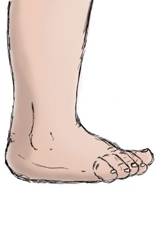
For better understanding of movements taking place in a foot it is best to read the article ANATOMY first. It will undoubtedly help you grasp the construction of a human foot. Because of your feet you can stand, walk, run, jump, ski, swim and do all sorts of activities. You touch the ground with them, feel the outlines and textures. All these things are possible thanks to 26 bones, which articulate in 33 joints reinforced with over 107 tendons and ligaments. They are moved by numerous muscles that work together in an amazing harmony to adapt to changing circumstances. Your foot will react differently depending on whether you’re walking on ice, or chasing a ball on a dense tennis court. When you jump- it adjusts and when you ski or skate- it does the same. It will work differently in all those scenarios. Regardless of the type of activity you’re attempting, it is all possible because of the mobility in joints of a foot. Joints are junctions of bones, that are connected to ligaments or tendons. They are protected by synovial capsules and synovial fluid and they can be moved by specific muscles.
JOINT
is a junction of two bones. They are connected via ligaments . The joint itself has two articular surfaces: convex and concave ( sometimes flat). Joint cavity is the space between them and it is filled with synovial fluid and optionally other structures (eg. ligaments, bursas, sesamoids, menisci) surrounded by the synovial capsule, which protects the joint from the environment.
LIGAMENTS
are strands of filamentous connective tissue made up of collagenous fibers with bundles protected by irregular sheath of connective tissue. Ligaments connect the BONES and reinforce the mobile junctions between them- joints.
TENDONS
are strands of filamentous connective tissue made up of very resilient collagenous fibers placed collaterally with bundles protected by thick irregular sheath of connective tissue. Tendons are silvery white colored. Tendons are extensions of MUSCLES and they connect them to BONES. They also forward the contraction of a muscle onto the bone.
MOVEMENT – MOBILITY AND GONIOMETER
When the muscles contract and release, their tendons move the bones they’re attached to and these bones move other bones linked to them via ligaments. This is just a summary of this complex mechanism. Movement is a very complicated process. This summary shows, that all the structures must collaborate, as they are communicating vessels: moving one moves the rest. Mobility is a feature of a foot, which is easily explained as ability to move. The range of motion (ROM) varies and is determined by the joints (their construction).

RANGE OF MOTION (ROM)
is a measurement of movement around a specific joint. Each joint can move in specific directions. The range of that movement is expressed in degrees. Range of motion for specific joints varies insignificantly depending on individual predisposition, age, gender. In general, ROM gets smaller as we age. Is ROM measured? Standard device used to measure ROM is called goniometer. It consists of two arms and a 0-360 scale. One arm is constant and shows 0 and the second one moves on the scale.

MULTIPLE MEASUREMENTS
measurements per second
The reconstruction of movement in three-dimensional recording systems means recreating the spatial orientation of the body movement. This is equivalent to finding the turnovers for each bone, on the basis of the data taken from special markers. Medicine has been interested in this technology from the very beginning. It can be used to diagnose diseases, plan treatment and track what progress this treatment brings. Thanks to the recorded images, it is possible to determine the trajectories of movement of the patient’s body parts, compare these movement paths with the accepted norms and determine whether there are any deviations from the norm. This allows us to improve the treatment or rehabilitation plan.
Since movement occurs in the joints, any soft structures involved in it can be both an aid and a restriction, reducing the range of motion or blocking motion altogether. In congenital clubfoot, there is a strong limitation of mobility (the range of motion is small or absent) due to a defect in the soft tissues, which causes problems with the alignment of the bones in relation to each other. The defect affects soft structures: ligaments, tendons, muscles. In them, a number of abnormalities appear, causing their pathology: shortening, calcification, fibrosis, which directly influences the bone structures, shifted relative to each other and slightly deformed. The pulling force of soft tissues is so great that it moves some bones relative to each other in certain directions.
MOTION IN THE ANKLE JOINT
Basically a joint, as you already know, is a connection of two bones with each other through a system of ligaments. The ankle joint is a complex structure consisting of many bones and soft tissues. The ankle joint is divided into:
- ankles mortise, also known as the talocrural joint (colloquially: ankle);
- ankles tarsus, otherwise known as the talocalcaneonavicular joint, is a complex, uniaxial, rotary joint.
ANKLES MORTISE
The ankles mortise otherwise known as the talocrural joint and colloquially as the ankle. It is composed of the ends of the tibia and fibula and the upper part of the talus bone. This is a typical hinge joint: the talus bone is partially located in the fork formed by the lower part of the tibia and fibula. The ankles mortise allows MOVEMENT OF THE FOOT UP (DORSIFLEXION) or DOWN (PLANTARFLEXION). While locking the foot on the ground, the joint allows the shin to tilt forward and backward. The range of these movements depends on many things: the training of the foot, age, collagen content in the soft tissues surrounding the bones.
NEUTRAL POSITION
Neutral position is the natural position of the foot when you stand on the ground. There is an angle of 90° between the foot and the calf (except it is actually 0° and it is taken as a reference point for measurements for dorsal and plantar flexion expressed in degrees).

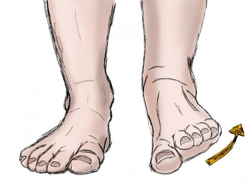
DORSIFLEXION
Dorsiflexion is the movement of the foot upwards towards the tibia. It is on average up to approx. 45°. In a normally functioning foot, dorsiflexion is inhibited by the tension of the posterior wall of the articular capsule and the calf muscles. Further more, the neck of the Talus eventually contacts the anterior edge of the articular surface of the tibia. It is referred to as “+” + the value of this movement. For example, “+ 15°” means the foot obtains 15° dorsiflexion.

PLANTARFLEXION
Plantarflexion is the movement of the foot downwards towards the ground and it is approximately 60°. In a normally functioning foot, plantarflexion is inhibited by the tension of the anterior wall of the joint capsule and the front muscles of the shin. In maximum deflection, the posterior process of the calcaneus rests against the posterior edge of the articular surface of the tibia. It is referred to as “-” + the value of this movement. For example, “-15°” means the foot has 15° plantarflexion.
THE ROLE OF THE ACHILLES TENDON IN THE DORSAL AND PLANTAR FLEXION MOVEMENT
The Achilles tendon, which is not directly related to the upper ankle joint, also contributes to the dorsiflexion and plantarflexion: its flexibility influences the range of motion performed. This means that if the Achilles tendon is fine and flexible, it will not restrict the upward and downward movement of the foot. Unfortunately, children with congenital clubfoot have thick, shortened and inflexible Achilles tendon, which makes dorsiflexion rather impossible. The foot is thus kept in a strong plantarflexion. The solution to this problem is the percutaneous Achilles tendon tenotomy.
ANKLES TARSUS
Ankles tarsus also known as the talocalcaneonavicular joint and is a complex, uniaxial, and pivot joint. Anatomically, two separate joints can be distinguished, devided by the talocalcaneal ligament:
- anterior ankle joint
- posterior ankle joint
Functionally they constitute one ankles tarsus. Due to its advanced structure, the ankles tarsus enables all rotational and torsional movements of the foot. The arrangement of these two joints: the ankles mortise and tarsus, gives the ankle the functional capabilities (freedom of movement) of the spherical joint, although in fact it is not.
POSTERIOR ANKLE JOINT
It is placed between the upper surface of the calcaneus and the lower surface of the talus bone. The main ligament of the joint is the talocalcaneal interosseous ligament. There are two movements in this joint:

INVERSION
Inversion is the movement of tarsal bone in which the undersurface of the bone moves towards the madian body plane. In inversion the lateral side of the foot touches the ground.
EVERSION
Eversion is the movement of tarsal bone in which undersurface of the bone moves outwards the madian body plane. In eversion, the medial side of foot touches the ground.
This joint also allows for various “variants” of positioning the heel relative to the ground, when standing, that is when the heel is fixed (locked) and weighted down. This applies to its three positions relative to the longitudinal axis of the foot, i.e. the axis running parallel to the ground.

NEUTRAL HEEL POSITION
Neutral heel position occurs when the heel is not deflected in any plane and receives the weight of the body with the entire surface of the calcaneal tuberosity. Both longitudinal arches of the foot are parallel to each other. The first and fifth metatarsal heads are evenly burdened, just as both sides of the foot (medial and lateral) rest evenly on the ground (the medial side has an arch, so it does not touch the ground with its entire length at the elevation).

HEEL VARUS
Heel varus is used for movements of inversion and adduction of the calcaneus.
In this position, the calcaneus flexes slightly plantarlly, causing a slight adduction of the forefoot and elevation of the medial longitudinal arch. Cavus begins.
The foot presses of the ground its lateral side.

HEEL VALGUS
Heel valgus is used for movements of eversion and abduction of the calcaneus.
In this position, the calcaneus flexes slightly dorsally, causing the foot to slightly abduct and lower the medial longitudinal arch of the foot. The foot presses of the ground its medial side.
ANTERIOR ANKLE JOINT (talonavicular joint)
It is placed between the anterior part (head) of talus and the concave surface of the navicular bone. It allows torsion:

NEUTRAL POSITION
Neutral position takes place when the long axis of the foot is parallel to the imaginary axis of the body, dividing it into the right and left sides.

ADDUCTION
Adduction is that movement of a tarsal bone in which the distal part of this bone moves towards the median body plane.

ABDUCTION
Abduction is that movement of a tarsal bone in which the distal part of this bone moves outwards the median body plane.
Everything seems relatively simple, if not for the fact that the foot has much greater mobility. These are linked movements. This means that one bone moves another one thanks to a system of ligaments, tendons and muscles. And this is how the foot can move in three planes, which you most likely are unaware of. Walking is the first stage in which the three-dimensional movement of the foot manifests itself. If you record your walk, slow it down and play it back almost frame by frame, it will turn out that your gait consists of many stages and each of them is necessary.

SUPINATION
Supination is a complex movement that occurs during the gait cycle. It is a natural movement which consists of inversion, plantar flexion and adduction. It occurs at the beginning of the gait cycle: first the floor touches the lateral side of the heel and the lateral side of the foot, just to do the opposite moments later. Supination gives the foot the stability it needs to balance. In supination, the calcaneus always moves into a varus position. If the foot is in supination, the lateral side of the foot is loaded more.

PRONATION
Pronation is a natural part of gait. It consists of eversion, dorsiflexion and abduction. It is the “middle” component of the gait cycle, in which the foot, after its first contact with the ground, rolls from the heel to the toe, leaning on the medial side to absorb shock by touching the terrain with a larger surface area than during supination. The foot is then very flexible and has great absorbent and adaptive properties. In pronation, the calcaneus always turns into a valgus position. If the foot is in pronation, the medial side of the foot is loaded more.
BLOCKED MOVEMENTS
Clubfoot is a three-dimensional deformity in which:
- the calcaneus is in inversion, varus, and plantar flexion (short Achilles tendon)
- the talus is medially displaced – adducted and in severe plantar flexion, which causes the calcaneus to be blocked underneath
- there is a very strong supination with a big cavus, and it would be no wonder – supination is a natural part of the foot activity – were it not for the fact that supination is constant and stable, preventing any other movement, effectively diminishing foots mobility.
Blocking the head of the talus and abducting the forefoot in supination (i.e. the original position) without touching the heel releases the blocked calcaneus and prevents deepening of the cavus. When the foot reaches its correct anatomical and functional function, the shortened Achilles tendon is subjected to tenotomy, which eliminates the last component of the defect – the equine position of the calcaneus (i.e. its strong plantar flexion caused by the Achilles tendon being too short).
UNDERSTANDING THE FUNCTIONAL ANATOMY OF THE TARSUS IS THE BASE THE CLUBFFOT TREATMENT IN PONSETI METHOD.
WITHOUT THIS FUNDAMENTAL KNOWLEDGE, TREATMENT IS BASED ON POOR CORRECTION, WHICH AFFECTS A SERIES OF PROBLEMS THAT COME UP “WITH TIME”.

SOURCES
RESEARCHES & STUDIES:
1. Ponseti I.V.: “Congenital Clubfoot. Fundamentals of treatment.” (2nd edition)
2. Michaud T.: “Human locomotion”
3. Earl J.: “Born to walk”
4. Basic Anatomy of the Foot
5. Marciniak W., Szulc A. et al.: “Wiktora Degi Ortopedia i Rehabilitacja”
6. Zukunft-Huber B.: “Dreidimensionale, manuelle Fußtherapie” (Edra Urban, Wroclaw 2015)
7. Supination and pronation
8. Ghanem I., Mosca V., Wicart P.: “Understanding the foot’s functional anatomy in physiological and pathological conditions: the calcaneopedal unit concept”
PHOTOS & GRAPHICS:
1. OptiTrack Systems
2. Other: own