Parents of infants born with clubfeet may be reassured that their baby, if otherwise normal, when treated by expert hands will have normal looking feet with normal function for all practical purposes. The well-treated clubfoot is no handicap and is fully compatible with a normal, active life.
DEFINITION
Congenital clubfoot (Latin congenitus talipes equinovarus (CTEV) or Latin pes equinovarus (PEV)) is the most common congenital deformity of the musculoskeletal system, characterized by complex deformation of the foot. It is not an embryonic deformation (i.e. a malformation) but a developmental one, because a properly developing foot turns into clubfoot in the second trimester of pregnancy, not just after fertilization or in the early embryonic stage, when organs develop (organogenesis). Congenital clubfoot has all the structures inside the foot (similar to a healthy foot) but at some point it begins to acquire the proper features of the clubfoot. The name „congenital” means that the defect is already present at birth and did not arise after birth, where the deformation is carried out on the originally healthy foot.
WHERE DOES THE NAME “PES EQUINOVARUS” COME FROM?
The name “pes equinovarus” is is a literal translation from Latin. The Latin adjective equīnus (equīna, equīnum) is related to the noun equus (equī), which means a horse. Why did the horse become the hero of this disadvantage? It is difficult to explain it unequivocally. Maybe because it is easy to explain one of the main components of the defect, on the example of the structure of the horse hind leg? It is significantly exposed.
The horse hind leg has a specific structure. The calcaneus and talus are located very high in relation to the metatarsal bones. A similar structure is shown by clubfoot, in which the calcaneus, by shortening the Achilles tendon, moves upwards, giving it a very similar position. A horse hoof is the equivalent of a human toe. Just as a horse stands on the hoof and the heel is very high, so also a child with clubfoot stands similarly: on the tip toes, and the heel is high and doesn’t touch the ground at all.
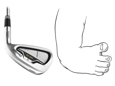
WHY “CLUBFOOT” ?
The deformed foot is very similar to the end of a golf club. In fact, when looking at a golf club and clubfoot, it’s hard not to see the similarities. This is how the English term for this defect was created: clubfoot (club – golf club, foot – foot). If you wanted to translate this term literally (as our Polish name for the defect has been translated), it would probably be a golf foot.
CHARACTERISTICS
Clubfoot is the result of several overlapping components. Only their joint appearance proves the correct diagnosis of the defect. It is a multifaceted and complex defect – it cannot be simplified, nor should it be underestimated.
In Latin, 4 letters define the “components” of a deformity:
C
cavus – excessive elevation of the foot medial longitudinal arch
A
adductus – foot adduction
V
varus – heel varus
E
equinus – equine position of the calcaneus
Incorrect ANATOMY OF THE CLUBFOOT, which is structurally advanced, is expressed in the external, very specific appearance of the foot:

an increased height of the vault of the foot
CAVUS (pes cavus), i.e. excessive elevation of the foot medial longitudinal arch. It refers to the increased height of the vault of the foot. The medial side of the foot is excessively elevated due to the strong contracted tendons, ligaments which thus position the metatarsal bones in the plantar flexion (especially the first metatarsal bone). It looks as if the foot is “broken” halfway through.

adduction
ADDUCTUS (pes adductus), or adduction. This is the limitation of the foot’s outward movement caused by shortened tendons and ligaments on the medial side the foot. Because of this, the foot moves inwards. This phenomenon can be observed by looking at the forefoot pointing towards the inside of the imaginary axis of the body, dividing it into the right and left sides.

heel varus
VARUS (pes verus), or varus. This is a feature associated with the calcaneus bone that tends inward so that the foot rests on its lateral side. This position of the calcaneus leads to its excessive inversion, which can be manifested almost by turning the sole of the foot upwards.

equine position of the calcaneus
EQUINUS (pes equinus), which is a equine position of the calcaneus. This is the most serious feature of this deformity. Too short the Achilles tendon causes the calcaneus to goes up and the forefoot is pointed downward (the foot is in a strong plantar flexion) so that the child can’t put the foot on the ground. It looks as if the baby is standing on tiptoe all the time.

thin calf
MUSCULAR ATROPHY of selected muscle groups in the calf. Muscle wasting (atrophy) in the calf on the side of the affected foot particularly affects the posterior and medial muscles of the calf. The triceps surae and the posterior tibialis muscles are fibrotic (excessive amount of connective tissue in relation to muscle tissue), are shorter and smaller, have a smaller volume. The difference in the size and volume of the calves is most noticeable in children with a unilateral defect.
TYPES OF CLUBFEET
Clubfeet can be divided into two main types, when it comes to their external appearance: typical/ standard and atypical. However, in each of these types of feet, certain subtypes can be distinguished related to the potential (because there are many hypotheses regarding its formation, and none of them has been confirmed so far) the source of their formation or certain factors that may cause them.
TYPICAL CLUBFEET

IDIOPATHIC – meaning “of unknown cause”. A child with this type of deformity has no other defects or diseases – the foot defect is not the result of another disease syndrome. You may also find the term isolated defect, which means the same thing. The child is healthy, “only” has a clubfoot. A variation of this type is THE POSITIONAL FOOT.

SYNDROMIC – the defect is associated with genetic mutations that lead to birth defects. So it is the result of a different syndrome. Such a foot often occurs in combination with a sequence, e.g. the Pierre-Robin sequence, a complex, e.g. diastrophic dysplasia or arthrogryposis (Latin multiplex congenita), a syndrome, e.g. Möbius syndrome, Larsen syndrome, Beckwith-Wiedemann syndrome, etc

NEUROGENIC – the defect is associated with other congenital malformations of a neurological basis, e.g. cerebral palsy (Latin paralysis cerebralis infantium), spina bifida (MC/MMC) with its varieties (Latin spina bifida), anchoring of the spinal cord or other types of paralysis. Such a foot is often more difficult to treat due to neurological connections, the direction of which can be difficult to predict.

TERATOGENIC – the defect occurs as part of disorders developed during pregnancy, e.g. as a result of taking drugs (amphetamines) or drugs that may have a strong effect on the fetus (most often they are strong antidepressants), smoking, drinking alcohol, poisoning with toxins.
NOT TYPICAL CLUBFEET
ATYPICAL AND COMPLEX FOOT are quite special types of clubfoot. The atypical clubfoot occurs only in the idiopathic form, while the complex clubfoot may apply to all of the previously mentioned types. Atypical and complex clubfoot, in addition to the standard features that you already know, has additional attributes: a characteristic deep crease above the heel and on the sole of the foot, the big toe is in the so-called hyperextension, the foot is in a very strong plantar flexion and the same equine position. The first factor differentiating both these feet is the time of their formation: the atypical foot can be recognized immediately after birth, before any treatment, while the complex foot is the result of a poor plaster casting technique with the addition of complications during the use of plaster casts: frequent sliding downe of plaster casts, leaving the plaster casts slipped on, swelling, casts deformation, e.g. when the heel is completely flattened by a poorly applied plaster cast. The atypical foot foot is relatively rare (in less than 5% of children with congenital clubfoot), the complex foot is more common.
GOOD TO KNOW
SOURCES
RESEARCHES & STUDIES:
1. Ponseti I.V.: “Congenital Clubfoot. Fundamentals of treatment.” (2nd edition)
2. Ponseti I.V., Smoley E.N.: “The Classic: Congenital Club Foot: The Results of Treatment.”
3. Ponseti I.V.: “To Parents of Children with Clubfeet.”
4. Pavone V., Chisari E. et al.: “The etiology of idiopathic congenital talipes equinovarus: a systematic review.”
5. Ippolito E., Gorgolini G.: “Clubfoot pathology in fetus and pathogenesis. A new pathogenetic theory based on pathology, imaging findings and biomechanics-a narrative review.”
6. Radler C.: “Foot Disorders of Newborns”
7. Grill F.: “Der Klumpfuß” (“Orthopäde 4/25”, str. 364-378)
PHOTOS & GRAPHICS:
1. Other: own and supporting people of good will