Parents of infants born with clubfeet may be reassured that their baby, if otherwise normal, when treated by expert hands will have normal looking feet with normal function for all practical purposes. The well-treated clubfoot is no handicap and is fully compatible with a normal, active life.
NONSTANDARD
Ponseti treatment is very effective and ensures that the feet are fully functional and in a normal appearance. This high efficiency and ease of treatment affects many feet. However, sometimes there are cases where the feet do not respond so easily to the standard treatment with this method. Such feet may include atypical and complex feet. The correctness of the treatment of atypical feet depends on the extensive knowledge and experience of the doctor who will meet them. Therefore, it is important to choose it very consciously. Similarly, in the treatment of complex clubfoot – medical experience plays the first violin, and it cannot be someone random, because the treatment requires some modifications made to the standard Ponseti method. But do not worry! These modifications are safe and effectively correct deformation without the need for extensive surgical correction or radically reduce the need for corrective surgery. Already in 1994. Dr. Vincent J. Turco discouraged any surgical operation on the feet, explaining that it becomes even more grotesque. He was also the first to classify them as “atypical clubfoot”. In 2006, Dr. Ignacio Ponseti along with colleagues informed about changes in the standard treatment protocol in order to be able to treat this type of feet as well. Treatment of atypical and complex feet using the Ponseti method involves not only redressing or plastering modifications, but also involves the use of a different type of foot abduction brace (suggest Mitchell brace) and its setting other than ‘standard’ positioning. All of this is important and will affect the health of these feet in the future.
REMINDER
The atypical foot is a deformity whose characteristic anomalies are formed in the prenatal period and manifest themselves shortly after birth, before any treatment. This is an idiopathic type of distortion. Complex clubfoot is a deformation resulting from inappropriate treatment of it at the stage of plastering – they are not applied correctly. Both types of feet are connected by a characteristic deep crease running across the foot, a deep crease above the heel and the first toe, which is in the so-called hyperextension to a greater or minor extent. The appearance of the feet and their individual features are presented in the article on ATYPICAL AND COMPLEX CLUBFOOT.
LITTLE BIG MODIFICATIONS
The modified Ponseti treatment protocol changes the way the manipulations are performed prior to plastering, and the process of plastering itself also differs from the classical approach. After the tenotomy, which is necessary, the plaster casts may be changed more often than usual. The protocol of using the foot abduction brace, its position and type are also changing. Correction requires precise find subtalar joint and a clear identification of the head of the talus. It is difficult to identify because it is very small: 5-7 mm, which is a significant disproportion compared to the doctor’s large thumb. It is also difficult to locate because it is less visible and protruding than the anterior tuberosity of the calcaneus, which is 1 mm below the head of the talus. If the doctor misidentifies the head of the talus and the thumb is too large to apply pressure to it, it will block the calcaneus, which should be able to move freely.
REDRESSION
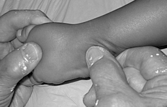
To be sure of the position of the talus head during stretching and when applying a plaster cast to the foot, the index finger should rest on the posterior side of the lateral malleolus, while the thumb of the same hand exerts counter pressure on the lateral side of the talus head rather than the very protruding anterior tuberosity of the calcaneus.
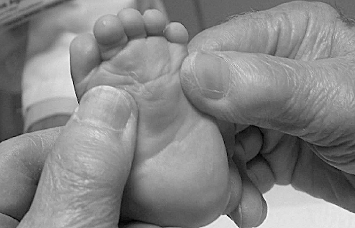
When the doctor reaches 30° abduction (not more!), he should change the way of redression, correcting the very strong plantar flexion of the metatarsals and, at the same time, the equine position of the calcaneus. He will place both index fingers on either side of the head of the talus and thumbs on the sole of the foot (exactly on the heads of the 1st and 5th metatarsal bones) and will ‘elevate’ all the metatarsal bones to a neutral level while allowing the blocked calcaneus to come out from underneath the talus. Redression is a manipulation that consists in stretching a tightly compressed plantar fascia and tendons in the sole of the foot and on the medial side. Only after all metatarsal bones have been brought out, it is possible to abduction them.
The alignment of the metatarsal bones (all bones) to the neutral level is very important – only after this procedure is performed it is possible to abduct the foot without creating additional deformations.
IMPORTANT
Additional attempts to correct the varus hindfoot (heel) by abducting the foot pushes the metatarsal and toes into additional plantar flexion and abduction, and a grotesque deformity is created. The lateral side of the foot acquires a transverse crease which gives it the appearance of being broken in midfoot.
You must not abduct the foot over 40° (approximately 30° is enough). If the doctor abduct it more, there is a high risk of increased deformation in the range of flexion and plantar metatarsal and toe metatarsal in hyperabduction Lisfranc joint.
PLASTER CASTS
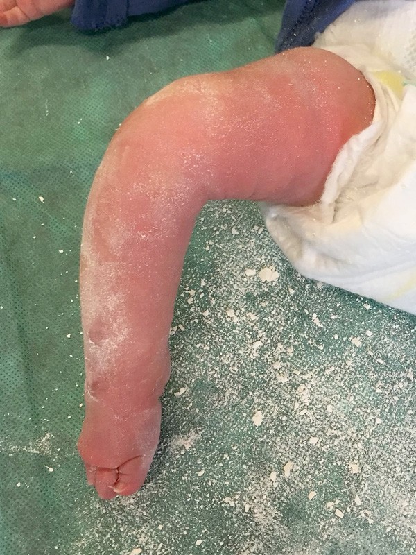
Complex clubfoot is formed by improper plastering – the plaster casts are poorly modelled, they often slip off, the foot and leg swells, it is bruised. Putting plaster cast on such a foot causes further deformations and fixes them. First, remove the slipping plaster cast. Secondly – change the doctor. Before re-plastering, it is necessary to wait until all the swelling has come off the leg and foot as putting on the plaster cast is pointless. Sometimes the break between plastering can be long. Long leg plaster cast is used, from the toes to the groin as in the standard treatment protocol Ponseti method!!! For the plaster cast not to slip off the chubby foot, the leg should be bent at the knee at an angle of 110°-120° (not 90°) during plastering. The plaster cast should be thinner under the knee and on the front of the ankle, and thicker on the front of the knee and on the thigh. The plaster cast must be well modelled so that it does not slip off. Especially around the heel and ankle. The toes should be clearly visible all over the place and slightly more exposed from the top. 6-8 plaster casts should be enough to correct the deformity (except in some more difficult cases, e.g. clubfoot in children with arthrogryposis).
TENOTOMY
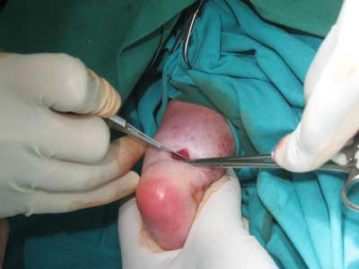
Tenotomy of the percutaneous Achilles tendon is practically necessary in the most of cases. It is performed when the doctor obtains a slight abduction of the foot (up to about 30°) and correction of the excessive plantar flexion of the metatarsal bone with the persistence of the equine (the equine position is uncorrected). A prior tenotomy may be needed if the cast slips off too often. The tendon is cut 1.5 cm above the crease on the heel. This distance prevents damage to the posterior tuberosity of the calcaneus, which is set very high. A minimum of 10° dorsiflexion is required to achieve correction. Unfortunately, this is not always achieved. Sometimes only 5° dorsiflexion is possible, and if this is the case, the post-tenotomy plaster casts may be changed weekly (up to 4 casts) until the result is (at least) 10° dorsiflexion and (maximum) 40° abduction.
FOOT ABDUCTION BRACE
The boots must be carefully made and fitted to prevent the feet from slipping out, and the FOOT ABDUCTION BRACE must be properly positioned. Such a comfort is provided using MITCHELL BRACE. The standard Denis Brown bar with lace-up boots is not enough for this type of foot: the foot will slide out very often, losing correction. It is also difficult to control the position of the heel in the boot, which is the key.
The initial position of the brace should be 30° – 40° of abduction (healthy foot: 30°- 40°). Later, when the foot takes proper shape, the abduction can reach 50° and should be increased gradually every few weeks to finally reach 60°.
EXCHANGE
It very often happens that a typical, regular clubfoot, because of poor plastering, turns into a compound foot. It is also often that many parents can hear that “the foot is difficult, non-corrective” and that it will require surgery, and it is best to operate it right away. This is not the way! Many doctors are also unaware that they create such a foot and they say that ‘it’s okay!’ and nothing to worry about. They often underestimate this type of deformity.
ZAMIANA ZWYKŁEJ STOPY KOŃSKO-SZPOTAWEJ W STOPĘ ZŁOŻONĄ NIE JEST “WINĄ” STOPY,
A ZŁEJ TECHNIKI GIPSOWANIA TYLKO I WYŁĄCZNIE!
Unfortunately, its further treatment is different from the standard Ponseti method and is associated with many difficulties in the future.
The risk of re-tenotomy increases significantly with this type of foot. Like the potential necessity of surgical intervention, which is 3 to 5%. However, even these surgical interventions should not be extensive and invasive.
RISK OF RELAPSE
HIGH RISK
Despite such a high risk of relapse, for children in the age group of 4-10 years, Ponseti treatment at the initial stage gives very good results, significantly reducing the need for invasive surgery. Sometimes when a relapse occurs, correct re-casting or a simple anterior tibialis tendon transfer (ATTT) surgery works well over invasive surgery like any release, including posteriormedial release that Dr. Vincent J. Turco said that they create an additional difficulty, which is the overcorrection of the foot manifested by a deep crease located on the lateral side of the foot. The earlier such an operation is performed, the deformation becomes deeper – the overcorrection is greater. Generally, there is no need for any surgery because the modified Ponseti method gives very good results, even in the event of relapse.
SOURCES
RESEARCHES & STUDIES:
1. Ponseti I.V. et al.: “Treatment of the Complex Idiopathic Clubfoot.”
2. Gupta A., Hakak A. et al.: “Atypical clubfoot: Early Identification and treatment by modification of standard Ponseti technique.”
3. Turco V.J.: “Recognition and Management of the Atypical Idiopathic Clubfoot”, Springer-Verlag New York, Inc. 1994
4. Radler C., Herzenberg J.E. et al.: “Ponseti versus traditional methods of casting for idiopathic clubfoot.”
5. Ponseti I.V., Morcuende J.A. et al.: “Radical reduction in the rate of extensive corrective surgery for clubfoot using the Ponseti method.”
PHOTOS & GRAPHICS:
1. Redressions: Ponseti I.V. et al.: “Treatment of the Complex Idiopathic Clubfoot.”
2. Tenotomy
3. Other: own