NOT COMPLETELY…
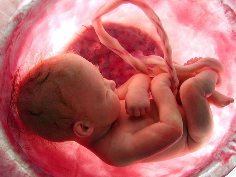
Perhaps you will come across a statement that your child’s defect is “positional because there was not enough space in the womb”. This belief is very common, inadequate and misleading. Lack of knowledge or ignorance of the deformity among the doctors, nurses and parents causes the statement to spring up like the proverbial mushrooms and thus reproduces a number of myths about the clubfoot. When we think about an “authentic“ it means structural clubfoot, it is formed when the baby still has a lot of space inside the uterus (the defect is created after the 12th week of pregnancy) and can move freely, unless severe oligohydramnios is diagnosed at such an early stage of pregnancy . Congenital and real clubfoot is not the result of the feet position inside the uterus. Although there was a theory blaming existence of some mechanical obstacle restricting the foot’s movement, hence causing it to be stuck in some position – it has been disproved as science and medical knowledge progressed and research developed.
Right now you are probably wondering: “Could there be a “nonauthentic” clubfoot?” The answer is… yes, it can. There are feet that look deceptively similar to clubfoot, but they are not actually clubfoot and the differences in diagnosis between the positional clubfoot and a mild degree of the clubfoot are very subtle and sometimes poorly defined, hence there is a lot of ambiguity in treatment as well as over-zealousness.
WHAT, WHEN AND HOW?
A positional clubfoot, also known as a habitual or postural is a very mild foot deformity, resembling clubfoot in appearance, but with a full range of motion. This means that the foot can move freely in any direction: there is no structural limitation and structural abnormality, which is present in congenital clubfoot. It is also not a foot classified as “soft-soft” (i.e. defined as the 1st grade in the Dimeglio Scoring System).
This deformity develops between 14 and 16 weeks of gestation, when the fetus still has a lot of space inside the uterus and when there is a large amount of amniotic fluid that allows it to move freely and without any obstrictions. It may be seen during a standard ultrasound scan, but only after the delivery will it be possible to determine exactly what type of defect it is and whether the original DIAGNOSIS was correct.
What causes positional clubfoot? When asked the above question by one of the parents, Dr. Ignacio Ponseti answered as follows:
(…) may be due to an anomaly in the myosin (the protein of the muscles in the posterior tibialis and gastrosoleus). This error in the myosin causes a contracture of the muscles and twists the foot in and down. (…) Incidentally the defect in the myosin is limited to the myosin of the flexor muscles of the foot. (…) After birth (…) the fetal myosin is replaced by normal myosin and the foot is normal for life.
RISK FACTORS
Among the risk factors contributing to the positional clubfood we can find the following:
- not enough space in the uterus, caused for example by oligohydramnios
- twin pregnancy
- some kind of mechanical obstruction (e.g. twisting of the umbilical cord)
- mother’s injuries during pregnancy arising from the pressure on the abdomen
It is difficult to say unequivocally whether these factors are primary or secondary. And is the problem with myosin in the muscles that are not working actively a result of their occurrence? Movement is extremely important for a fetus. It keeps its body in good shape. Cessation of movement causes many problems, such as contractures. Movement also allows for the proper development of muscles: if there are no restrictions on their development as a result of an obstruction – muscle fibers can work and develop.

CHARACTERISTICS
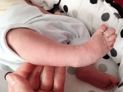
The positional clubfoot is of normal size and slender shape. There is no difference in the length and width of the feet as noted in children with congenital clubfoot with a structural basis (i.e. exhibiting changes existing in the structure of bones and soft tissues). The bones inside the foot are not shifted against one another. Their shape remains unaltered. The Achilles tendon in the positional clubfoot is of adequate length and width and is not fibrotic. Its functions are preserved. This is evidenced not only by the possibility of obtaining a good dorsiflexion of the foot, but also the skin folds above the heel are the same as in a healthy foot. Likewise, the calf muscles are not fibrotic and smaller (hence the circumference of the calf is practically the same as in a healthy leg), and the potential “error” in myosin does not reflect in the characteristic PATOANATOMY OF THE CLUBFOOT.
RANGE OF MOTION
The positional clubfoot can be moved freely in any direction. It is passively manipulative, which means that during the examination the person who moves the foot does not put much effort into this movement, and the child moves the foot consciously using the strength of its muscles. Positional clubfoot exhibits excellent MOBILITY:
- it can be easily abducted (abduction is a movement outward from the imaginary axis of our body, dividing it into the right and left sides)
- it can be flexted upward (dorsally): the degree of flexion will vary, but it is greater than 0° (0° being the foot placed on the ground, i.e. reaching an angle of 90° between the calf and the sole of the foot)
- the foot has a full range of motion both in inversion and eversion, supination and pronation
TREATMENT
Generally, the postitional clubfoot does not require a treatment, as it “passes on its own” shortly after birth, after 3 months of age, or when the baby starts to stand up and walk, weight-bearing. If treatment has to be applied, the foot responds easily to it, and the effectiveness of conservative treatment (stretching and massages) is high. Post-correction foot development is normal. There is no recurrence of the defect. However, some cases fall somewhere between a hard, but still positional, defect and a mild, idiopathic clubfoot. They should be observed until the end of the second year of life. Since the habitual defect is relatively light and the line between it and a slight clubfoot (Dimeglio 1st) is very thin, some doctors believe that nothing should be done about it and underestimate the problem. It is very important to be able to distinguish between these two defects, as the treatment is very different in both of the cases. Sometimes a slightly stronger positional defect requires the use of cast, as well as the use of a foot abduction brace for some period of time.
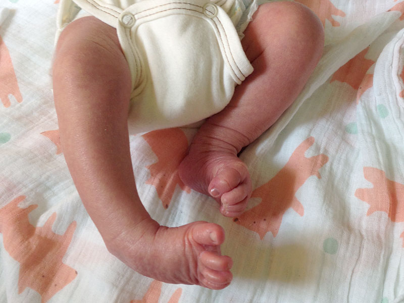
Positional clubfoot is a molding deformity which resembles clubfoot. The foot is in mild equinus, in adduction and is rotated inwards. However, the foot is passively fully flexible with dorsiflexion well above neural and no sign of the rigid deformities seen in true clubfoot. Positional clubfoot usually responds to stretching and massage. However, some cases are somewhere between severe positional clubfoot and mild idiopathic clubfoot and should be followed to at least two years of age. Whenever a cast is needed for correction, abduction bracing as for clubfoot should be performed for a limited amount of time.

SOURCES
RESEARCHES & STUDIES:
1. Radler C.: “Foot Disorders of Newborns”
2. Anand A., Sala D.A.: “Clubfoot: Etiology and treatment.”
3. Furdon S.A., Donlon C.R.: “Examination of the newborn foot: positional and structural abnormalities.”
PHOTOS & MOVIES:
1. Baby in womb
2. Photo under quote (bottom of the page): Radler C.: “Foot Disorders of Newborns”
3. Other: a support of the people of good will