The human foot is a masterpiece of engineering and a work of art.
HISTORICAL DIFFERENCIES
It is hard to disagree with the above statement of the master. Da Vinci, as a keen observer and inquisitive researcher, noticed things that we often do not think about anymore.
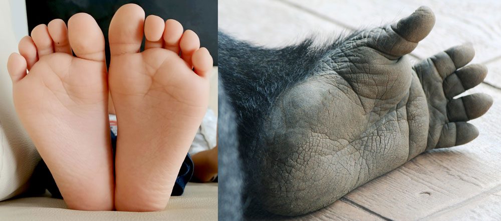
The foot is an organ found only in primates; however, only in man does the foot have such a specific structure, which allows him to walk continuously in an upright and bipedal position without having to assume a quadripedal position. The change in man’s lifestyle, the conditions in which he lived, and the work he did required the evolution of the foot, the effects of which we are benefiting from today, because it is the upright position that has influenced the shape of the foot today.
Unlike the ape-like primates, the human foot no longer has an opposable big toe, which is an aid when moving through tree branches (the foot in apes has an analogous arrangement to the hand – it is capable of grasping). The reduction of the opposing toe in favor of the lengthening of the first metatarsal bone has resulted in man being able to stand and walk upright freely and stably, and the big toe is one of the supports that makes this way of moving possible (the human foot has 3 points of support).
ANATOMY

The foot is the distal part of the lower limb located just below the ankle.
The basis of its structure and activity is:
- at least 26 bones (1/4 of our body bones are in the one foot)
- at least 33 joints
- at least 107 tendons and ligaments, the movement of which causes the work of numerous muscles.
All of the above structures are placed in one foot and each foot is like that, which, however, is quite small area. This makes its construction very complicated. It can be said to be one of the most advanced organs in our body.
The surface of the foot is covered with skin in which there are sweat glands (no sebaceous glands), numerous nerve endings (there are over 200,000 of them in the sole of the foot) and small blood vessels. The fingertips protect the keratin nails, preventing them from being injured.
TRIANGLE
Viewed from the sole, a properly formed foot is shaped like a triangle: wide at the toes and narrow at the heel. This is the shape children’s feet retain for a long time, until they start wearing the wrong footwear. The three points of support give it just this triangular appearance.
The foot touches the ground with the tuberosity of the calcaneus and the heads of metatarsal bones which lie in one line and at the same height. This means that the concept of a transverse arch at the heads of metatarsal bones of the foot doesn’t exist. Looking from the same perspective, i.e. in the frontal plane, moving towards the heel, there is the transverse arch passes through the three cuneiform bones and the cuboid bone.
Looking at the foot from the sides: from the medial side of the foot the medial longitudinal arch is formed and it runs from the calcaneus through the navicular bone, the first cuneiform bone to the head of the first metatarsal bone. The top of this arch is the navicular bone (about 2.5 cm above the ground).
From the lateral side – the lateral longitudinal arch is formed and it connects the calcaneus with the head of the fifth metatarsal bone passing through the cuboid bone.
Such a “triangular” structure of the foot enables not only stable standing but also carrying heavy loads and shock absorption during walking, running and other activities.

SIGNIFICANT DIFFERENCES
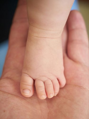
At this point, it is advisable to make a distinction in terms of the development of the foot and its anatomy: the foot of child it is not the same like the foot of adult. This is a very important distinction as a child’s foot is different from that of an adult. These differences are not only in the external appearance (which is obvious), but mainly in its internal structure. A child foot is not a miniature of an adult foot.
Toddler’s foot is wider in the toes, narrower in the heel, as it is not yet shaped by bad shoes, mass, and activity (or lack thereof).
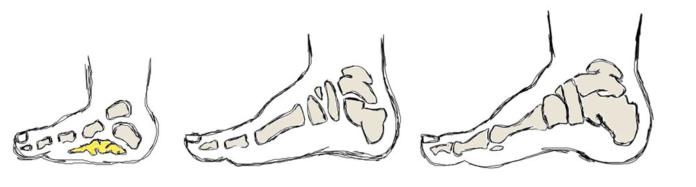
There are many cartilage structures inside the foot because the bones do not ossify quickly and evenly. The bones are mostly cartilaginous (made of cartilage tissue) and thus they are very soft and delicate, susceptible to deformation and are able to adapt to many unfavorable shapes (e.g. too small shoes). They do not grow evenly, and the process of their ossification takes several years. This unevenness can be seen on the example of shoes, when the foot quickly “skips” the size of the shoes: let’s say size 24 is too small after two months of use, but the child “takes” into size 26 and not 25.
OSSIFICATION - WHAT IS IT?
Tendons and ligaments often have a higher collagen content: they are very flexible and plastic, their adaptability to conditions is very large and the range of motion in the joints is also greater than in an adult human with an already formed foot. Tendons and ligaments are also weak and vulnerable at the same time.
The medial longitudinal arch and the whole sole of the foot is filled with a thick layer of adipose tissue. It seems that the child has no longitudinal arch because it puts the whole foot on the ground. Many parents are unconvinced that their children have flat feet. The thick layer of adipose tissue also acts as a great insulator: although children run barefoot on various surfaces and the foot may seem “icy”, in reality it is not, because it is the “fat pad” that protects the foot from freezing.
LONG-TERM FORMATION
From the prenatal period until around the age of five, a child’s foot undergoes a number of amazing changes to be able to ultimately fulfill its functions. Of course, the fifth year of life is not the end of the transformation because it takes on its own shape and features for a long time. At some point, the bones finally form and harden, tendons, ligaments, and muscles strengthen, and the adipose tissue of the medial arch and sole disappears. Generally, the human foot is formed around the age of 12-14, but is finally “finished” around the age of 17. It is worth not to disturb the feet in their development – they are the pillar of our body.
Consciously, the foot anatomy will be discussed on the example of the adult human foot skeleton. This is due to the fact that such a foot is already well formed: all bones are “finished” as to their shape, and they are strong and hard. Tendons, ligaments and muscles are well formed.
The traditional split divides the foot into 3 parts:

FOREFOOT

MIDFOOT

HINDFOOT
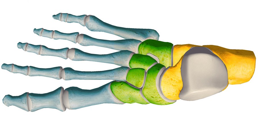
The hindfoot and midfoot form the tarsus, and this section will focus on discussing this complex because it is directly related to the defect – the clubfoot.

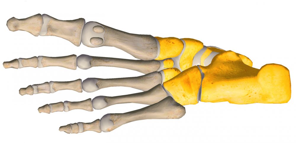
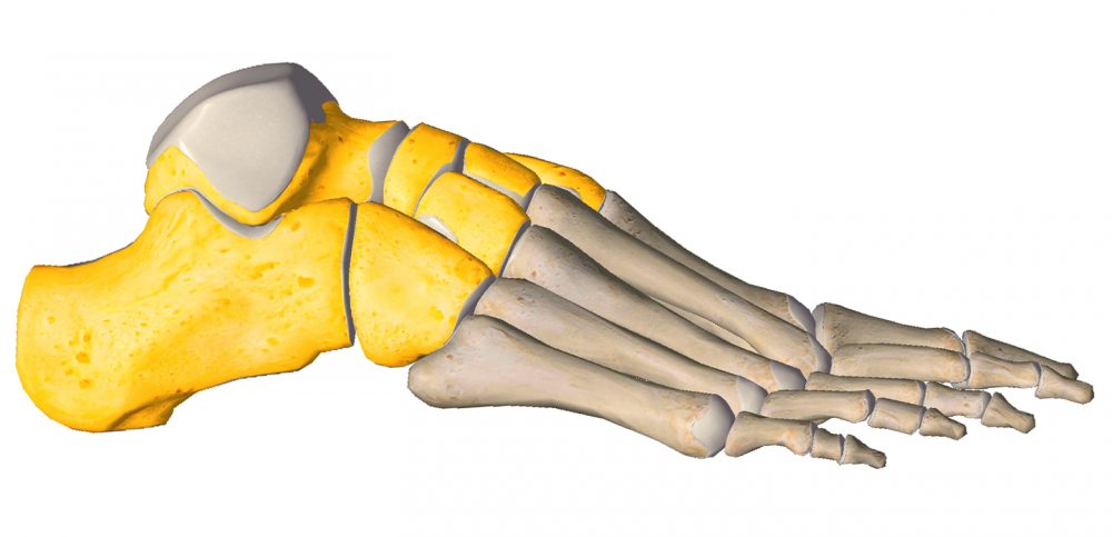
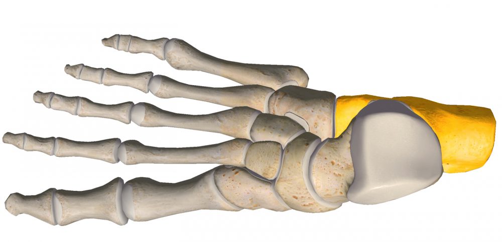
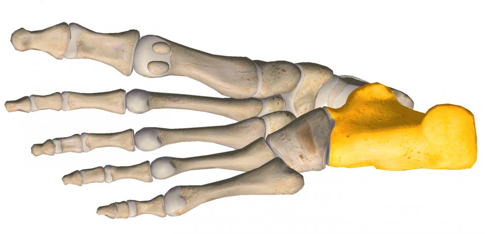
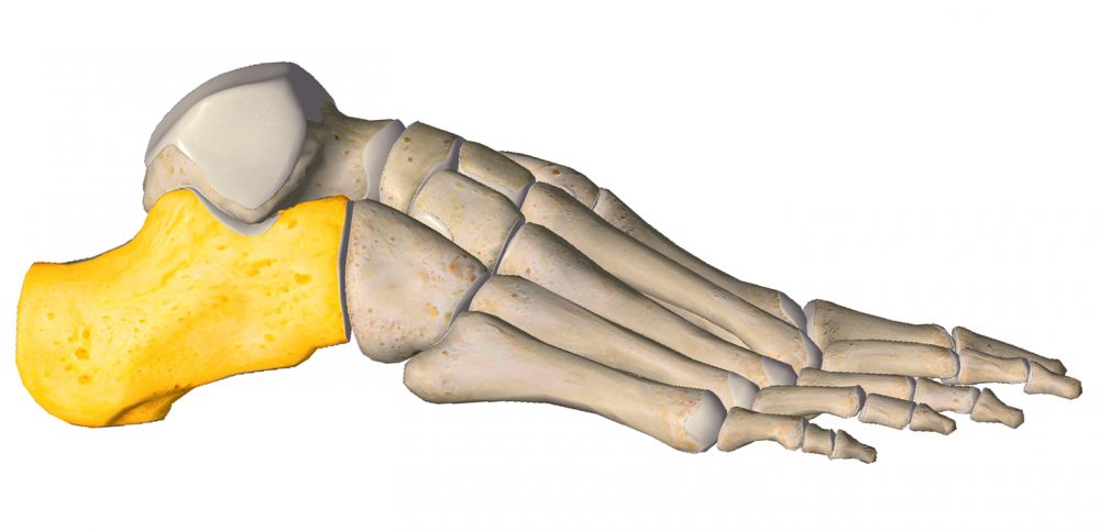
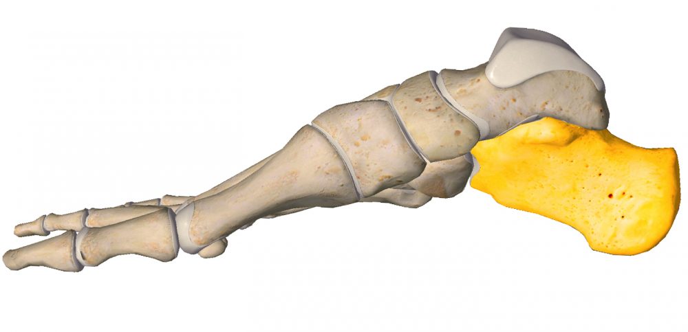
CALCANEUS
CALCANEUS (Latin calcaneus) or heel bone is the largest of the tarsal bones and the largest bone of the foot. It is located in its posterior lower part under the ankle bone. The calcaneus articulates on top with the ankle bone at the subtalar joint and on the front it articulates with the cuboid bone at the calcaneocuboid joint.
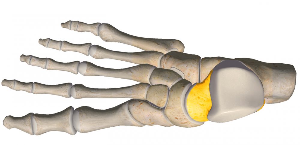
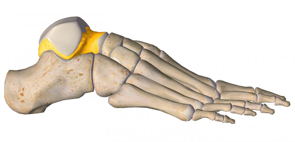
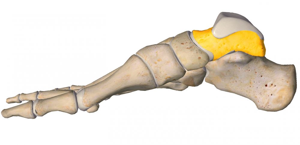
TALUS
TALUS (Latiun talus, astragalus) is a one of tarsus bone. It is located between the tibia (upside) and calcaneus (downside), betweent lateral and medial malleouls. Facing anteriorly, the head carries the articulate surface of the navicular bone, and the neck, the roughened area between the body and the head, has small vascular channels. Because of the connections between these bones, the ankle bone transfers all the weight-bearing of the body to the foot. No muscles attach to it.
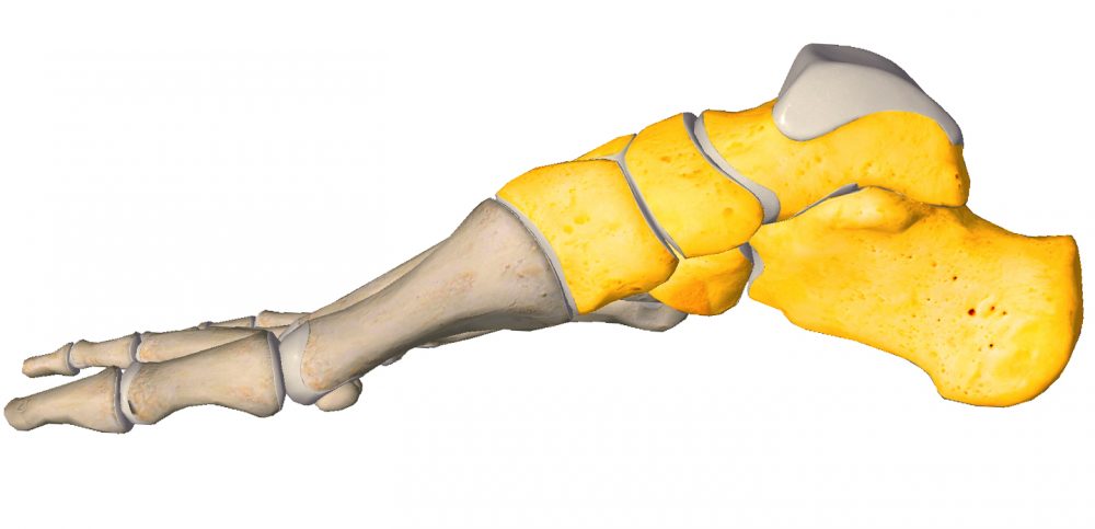
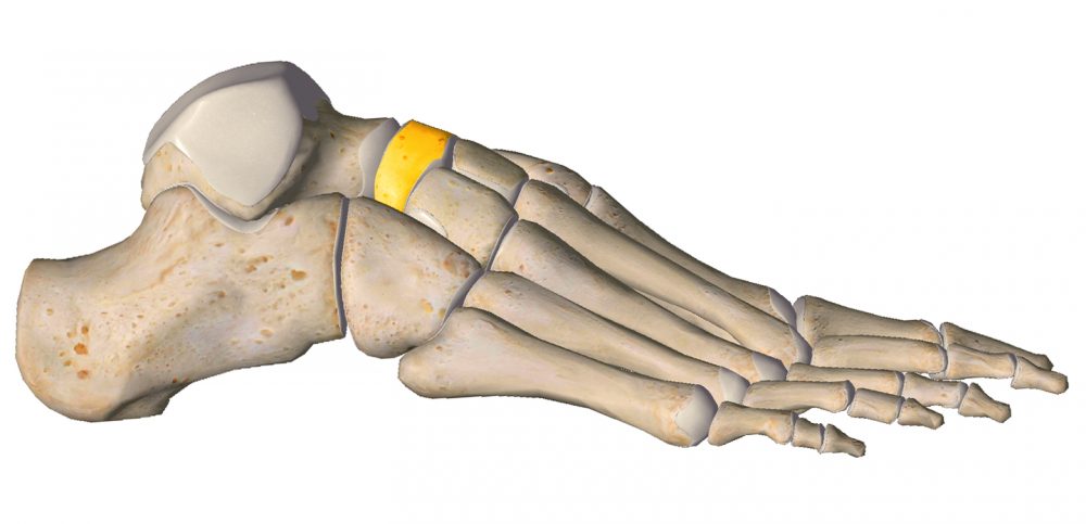
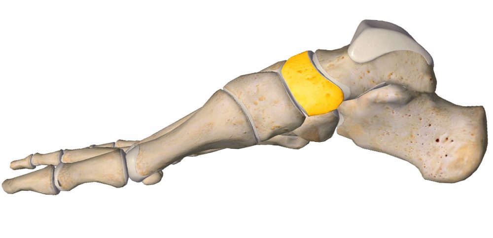
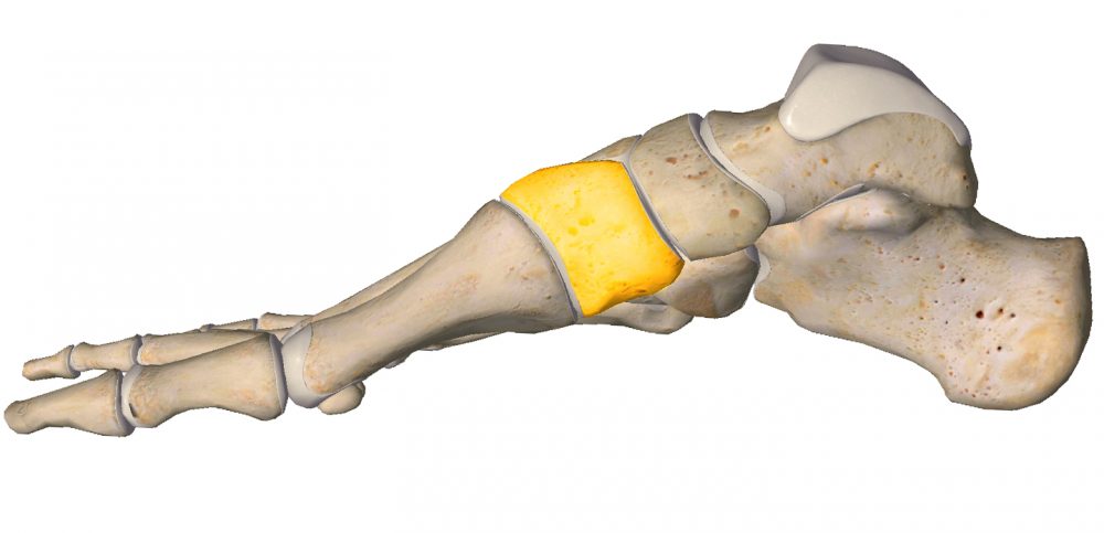
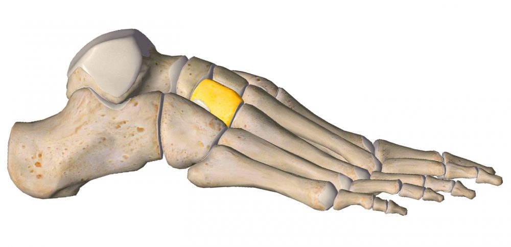
NAVICULAR
NAVICULAR (Latin os naviculare) is a part of tarsus. On the medial surface, it has a prominent tuberosity (Latin tuberositas ossis navicularis) – a control point when examining the transverse tarsal joint. On the anterior surface, it has three fields for connection to the three cuneiform bones. The lateral surface has a non-permanent articular surface for the cuboid bone, while the posterior surface has a non-permanent articular surface for the ankle bone.
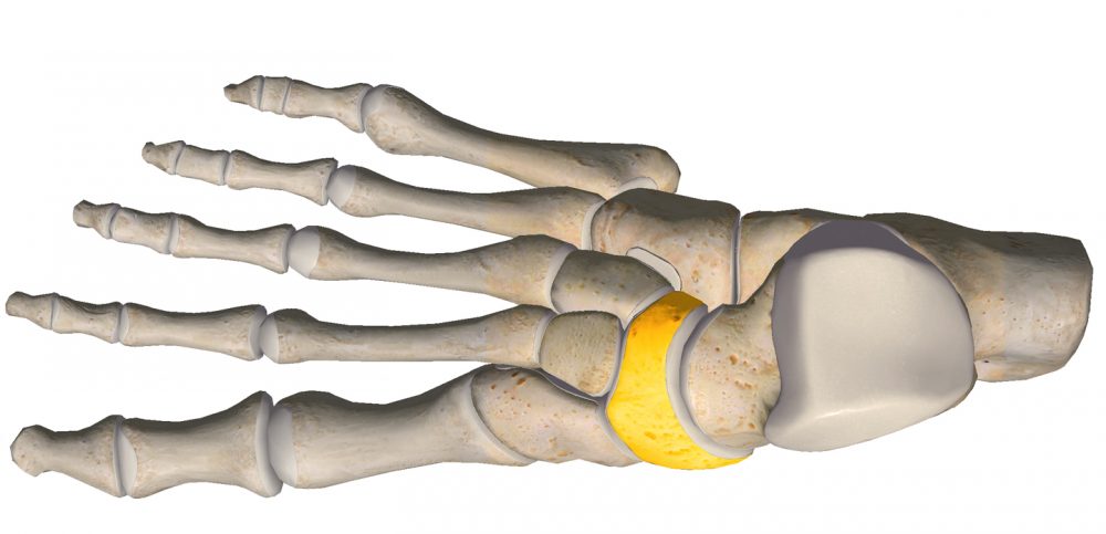
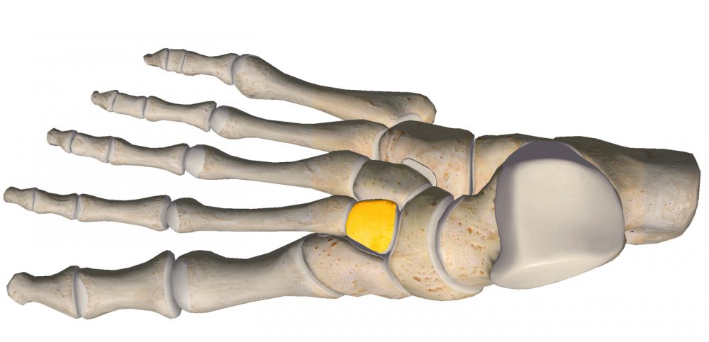
CUBOID
CUBOID (Latin os cuboideum) has an uneven cube shape and is placed anteriorly from the calcaneus on the lateral side of the tarsus. Its medial surface is longer than its lateral surface.
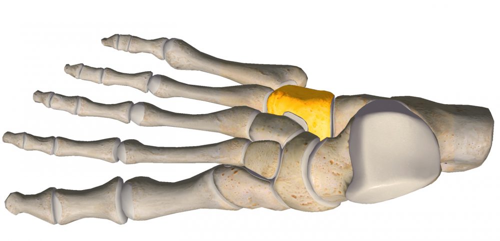


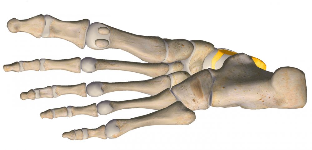
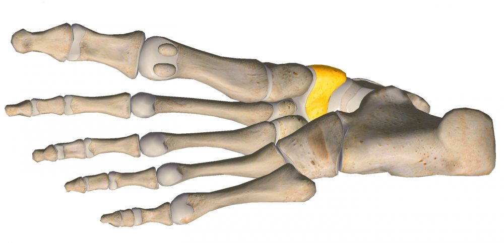
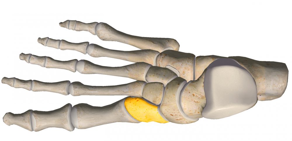
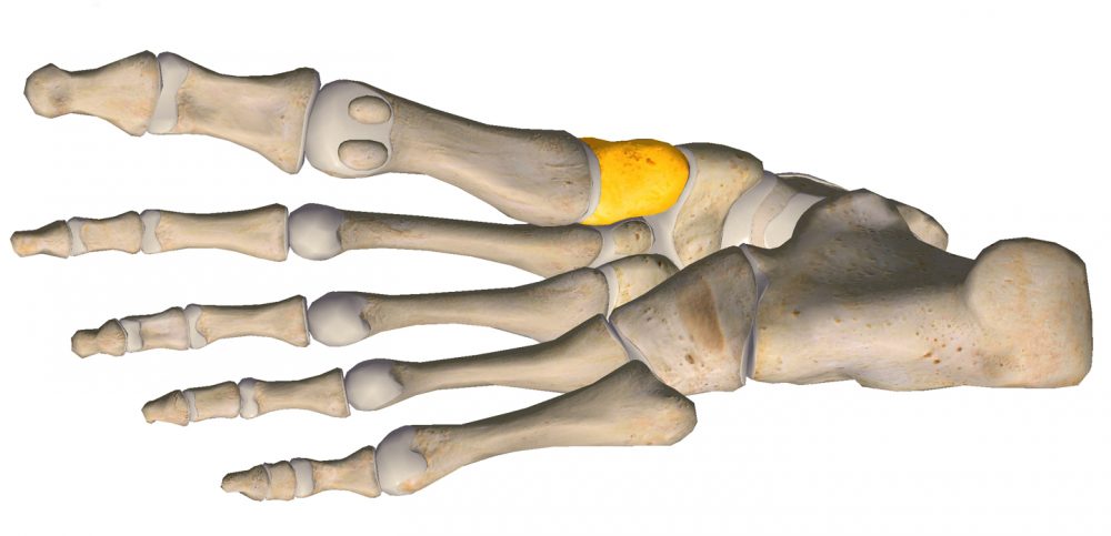
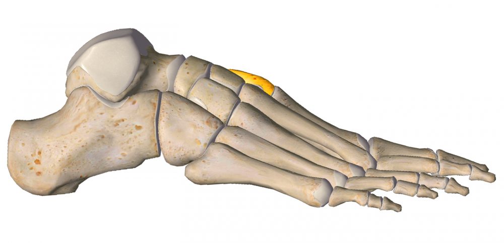
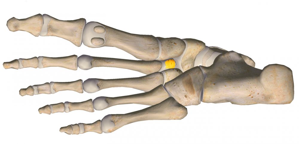
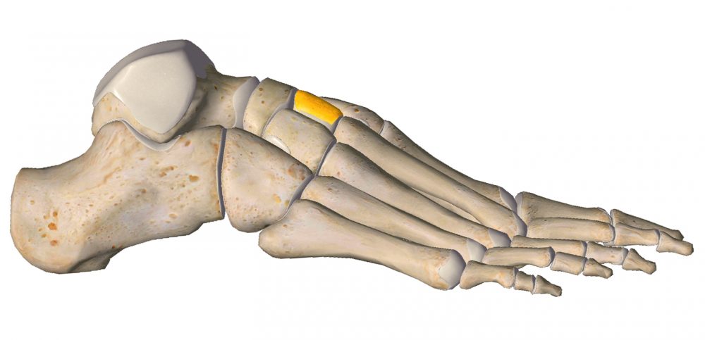
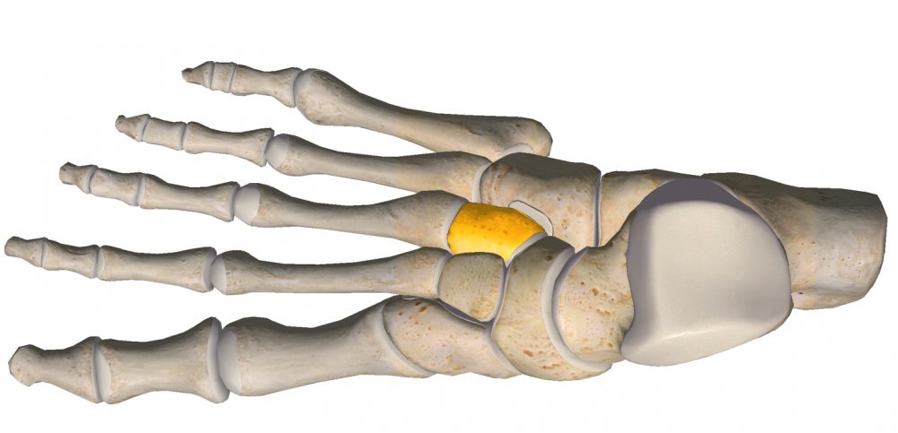
CUNEIFORM BONES
CUNEIFORM BONES/ CUNEIFORMS (Latin ossa cuneiformia) are located between the navicular bone and the first, second and third metatarsal bones and are medial to the cuboid bone. They are wedge-shaped. There are 3 cuneiform bones: medial, intermediate and lateral.


MULTIFUNCTIONALITY
The foot is one of the organs that play key roles in making our lives and functions easier.
LOCOMOTION
The foot is a locomotor organ.
It allows you to move wherever and however you need to: walk, run, jump.
SPAR OF YOUR BODY
It is the spar of your body.
Body weight – bones, muscles, organs, etc., is ultimately supported by the foot while standing, walking and running.
DO YOU KNOW...
SUPPORT
It is the support of the body.
This means that thanks to the three points of support mentioned earlier, i.e. the “triangle” stretched between the tuberosity of the calcaneus, the heads of the first (I) and fifth (V) metatarsal bones, and thanks to the complement of the triangle by ligaments, tendons and muscles between these points , specific “bridges” are formed. They allow you to stand steadily regardless of the surface.

SHOCK-ABSORBER
It is a shock-absorber.
Thanks to its advanced structure (“a masterpiece of engineering”), MOVEMENTS occurring in the joints of the foot allow for propulsion of the foot while walking, running, jumping, sliding, … (enter any variations that come to your mind in connection with the foot here) during sudden, while allowing you to adapt to the terrain.
This is important because the foot works in various planes and its adaptive properties affect the stability of the body and the transfer of large and variable loads in order to protect the joints from excessive stress leading to injury and you from falling.

TRANSMITTER
It is a transmitter.
Thanks to your feet, you recognize the terrain – not only its shape but also its temperature and structure. This is thanks to numerous nerve endings of which there are over 200,000 in the sole of each foot and numerous blood vessels. It is the contact of the foot with the ground that helps us determine whether you are walking on wet grass or hot sand, whether the terrain is sloping or dipping. Such sensory experiences are extremely enriching in terms of overall human development and cannot be underestimated, especially in children.
DISCOVER!
TAKE CARE!
The entire correct posture of the body begins with the foot. It is necessary to take good care of this locomotor organ to avoid complications in the future that you may not be aware of now. Many orthopaedic problems of children are connected with abnormal formation of the foot: bad shoes, too much body weight, lack of activity, forceful support of the development by not (or limiting) lying on the stomach, placing the child by the furniture when it is not ready, leading it “by the hands” and facilitating its functioning in many aspects: beds, rocking-toys, walkers, soft mats, giving toys. Such “support” does not bring the intended results. A child has to learn to use its feet because each stage of development is necessary for its little feet to be strong and healthy.
Nature has designed us wisely – do not disturb her, and she will manage!
SOURCES
RESEARCHES & STUDIES:
1. Barisch-Fritz B., Mauch M.: “Foot development in childhood and adolescence.”
2. Sarrafian S.K., Kelikian A.S.: “Development of the Foot and Ankle.”
3. Mosca V., Ghanem I., Seringe R.: “Understanding the foot’s functional anatomy in physiological and pathological conditions: the calcaneopedal unit concept.”
4. Foot
5. Tarsus
6. Foot of child
7. Developmental of child’s foot
8. Zukunft-Huber B.: “Der kleine Fuß ganz groß. Die dreidimensionale, manuele Fusstherapie auf neurophysiologischer Grundlage.“ (Edra Urban, Wroclaw 2015)
PHOTOS & GRAPHICS:
1. Gorilla’s foot
2. Screenshots: Essential Anatomy 3 / 3D4Medical: own
3. Other: own