WHAT IS THE ACHILLES TENDON?
Achilles tendon is heel cord, also known as the calcaneal tendon. The Achilles tendon connects the calf muscles to the calcaneal tuberosity. However, its structure is more complicated than it seems. Interestingly, the Achilles tendon is the thickest and strongest tendon in your body, and at the same time it is at the highest risk of injury. Such a paradox.
MYTHOLOGICAL ROOTS
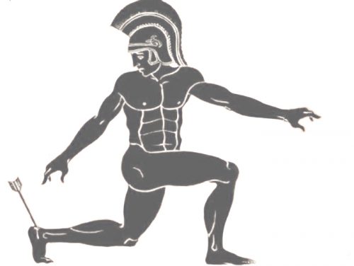
Perhaps you are wondering why the tendon is called a name of a mythical hero? There are two versions available for information. The first of them says that Achilles was immersed in the waters of Styx as a child by his mother – the sea goddess, the nymph Tethys. Thanks to this, his body was to be immortal and resistant to any blows. Unfortunately, the heel that his mother held him by during the bath was not submerged, which made its his weakest point. The second version says that during the Trojan War, Achilles was stabbed in the heel by a poisoned arrow of Paris and was thus killed.
STRUCTURE
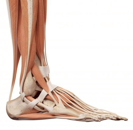
Achilles tendon, although it seems simple, is one of the most interesting tendons in terms of anatomical structure. It is 15 cm long on average and up to 2.5 cm in diameter in cross section. It is not homogeneous in width and thickness: high it is flat, thinner and wide, then it becomes oval and narrow (approx. 4 cm above the attachment to the calcaneus), then widens again a bit and flattens, sticking to it and looking like a fan. This insertion may appear at different heights and cover a different surface, which is an individual feature of a given person. It is made of collagen fibers, but they are not very stretchy. The tendon is relatively poorly supplied with blood and, at a certain height, poorly innervated, which contributes to its long healing time, e.g. in the event of a rupture injury.
MUSCLES
It consists of the distal fibers of the gastrocnemius muscle and the soleus muscle, which is slightly highter than the former. These two muscles make up the triceps surae. In 93% of cases, the plantaris muscle that runs along the lateral edge may be the companion muscle that supports the Achilles tendon. On this basis, it can be said that the calcaneus tendon consists of two layers that merge into one at some point. This is partially true. At a certain height, the ends of the gastrocnemius muscle join the ends of the soleus muscle through the aponeurosis, which fuse together.
APONEUROSIS

Gastrocnemius muscle is a superficial muscle that is in the back part of the lower leg of humans. It is two-headed muscle: lateral and medial. With the soleus muscle, it forms the triceps surae muscle of the calf, which in turn forms the Achilles tendon. The main function of the gastrocnemius muscle is plantar flexion of the foot at the ankle joint and leg flexion at the knee joint. It also corresponds to fast leg movements: jumping, running fast because it is made of fast twitch fibers, unlike the soleus muscle. The soleus muscle owes its name to … fish. Sola is a flat and wide sea fish, the shape of which is similar to the soleus muscle. Like the gastrocnemius muscle, this one is also a superficial muscle hidden behind the gastrocnemius muscle. Together with it, it forms the triceps surae muscle of the calf and the Achilles tendon. Its main function is plantar flexion of the foot. It also plays a key role in maintaining your standing posture: if it weren’t for the constant tension in standing and pulling, your body would fall forward. Its long-term work is related to its structure – they are slow twitch muscle fibers. In the standing position, it is responsible for pumping blood to the heart.

Plantaris muscle is a superficial muscle that runs between the two mentioned above. It is absent in 8-12% of the population. It is not part of the Achilles tendon, but supports it significantly. The plantaris acts to weakly plantar flex the ankle joint and flex the knee joint.
The plantaris muscle may also provide proprioceptive feedback information to the central nervous system regarding the position of the foot. Its motor function is so minimal that its long tendon can readily be harvested for reconstruction elsewhere.
BURSAE
BURSA
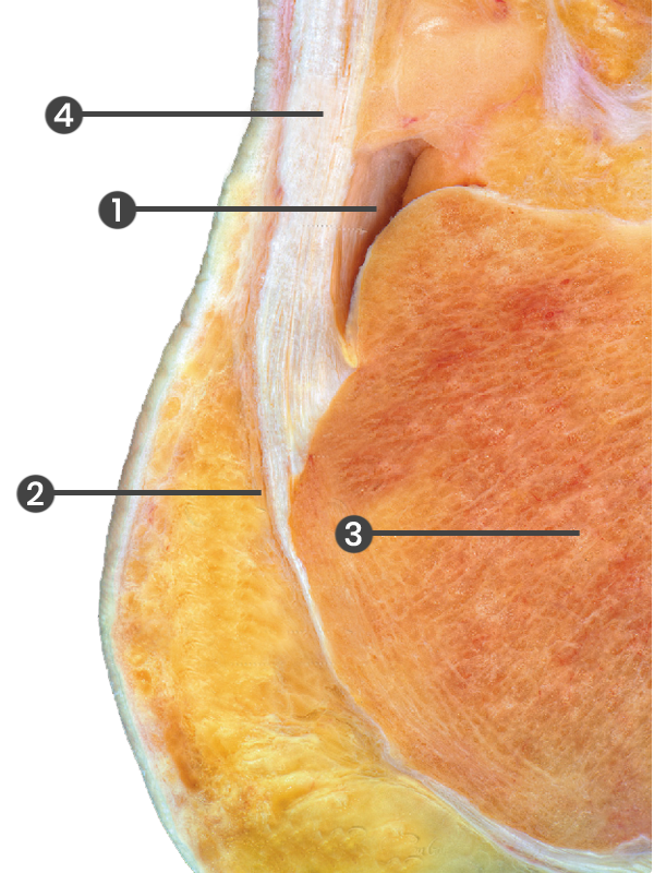
Between the anterior part of the tendon and its insertion, there is the bursa of calcaneal tendon. It prevents excessive friction. However, this is not the only bursa in this place – the second one is between the tendon attachment to the calcaneus and the skin and is called the subcutaneous calcaneal bursa. It protects the tendon attachment. As the tendon works, the bursae move between the layers of tissue. This can be seen especially in the case of the Achilles tendon bursa, which is hidden between the tendon and the calcaneus. When you bend your foot up (dorsiflexion), the bursa moves upward slightly. When you bend your foot downward (plantarflexion), the bursa moves downward. The bursa also has its protection in the form of adipose tissue located in the area of the Achilles tendon insertion, creating a specific, triangular shape. This is called Karger fat pad, which protects the bursa against excessive pressure and from moving sideways.
1. the bursa of calcaneal tendon | 2. the subcutaneous calcaneal bursa | 3. the calcaneus | 4. the Achilles tendon
KARGER’S FAT PAD
KARGER'S FAT PAD
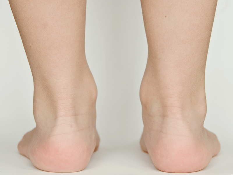
Have you ever wondered why the sides of the Achilles tendon are empty and can be caught with your fingers through the skin? This is most evident when looking at the foot from behind. It looks like there is empty space there, but it is not!
The main function of Karger’s fat pad is minimalization of the pressure in the subcutaneous calcaneal bursa during the bend the foot up and the protection of the joint capsule against overload and breakage. This structure also prevents other collateral structures from shifting, which minimizes injuries.

TORSIONED
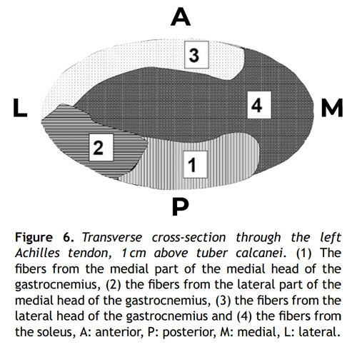
The Achilles tendon can be said to have a four-bundle structure.This means that the tendon is made up of four tendon bundles, which are the ends of the muscles: three of them come from the gastrocnemius muscle and one from the soleus muscle. Importantly, these bundles are not arranged parallel to each other, but are twisted in a characteristic way, which affects the function of the tendon. How are they twisted? Differently. Because it is not the same for every human being, although there are certain types of fiber twist. They reach 90° at the insertion to the calcaneus. This twisting of the bundles gives the tendon high strength, as it is able to take a load 3.9 times greater than the body weight while walking and 7.7 times greater than the body weight while running. Unfortunately, such a structure also affects his injuries, which are sometimes difficult to heal and recovalence takes a long time.


FUNCTIONS
The intricate and advanced construction of the Achilles tendon allows you to stand, walk, run, jump, twist your feet to the side, load them on the lateral and medial edges. The main function of the tendon, however, is to transmit to the foot (through the calcaneus) the force generated by the calf muscles, and the function of these in turn is plantar flexion of the foot. So the Achilles tendon is actively involved in bending the foot down. The calf muscles then tighten, pulling the tendon, which pulls the heel bone up. 93% of this force goes to the tendon. For any FOOT MOVEMENT to occur, the muscle strength is transmitted through the tendons to the bones. This is an amazing cooperation.


LIKE A SPRING…
The calcaneal tendon is involved in almost every physical activity of the foot. Plantar flexion movement is needed with each step as you move. The tendon stores some kind of energy that is spent on foot activity. This is called elastic energy. The Achilles tendon is like a spring that, while stretching, must “pull” to release the stored energy, and this is precisely what happens during the plantar flexion: energy is released. However, in order for the tendon to accumulate energy and give it back, it is necessary that it is sufficiently stiff, i.e. resistant to high forces, and at the same time flexible, so that it returns to its shape and elastic to release energy.
STIFFNESS
FLEXIBILITY
Both of these features are necessary for the tendons. Thanks to their stiffness, the tendons do not break, they also maintain some stabilization between the muscle and the bone. However, their flexibility means that they do not deform, changing the structure, which would translate into their functions. The lower the stiffness, the greater the flexibility and elasticity to be able to accumulate energy more efficiently and then release it. However, too much flexibility and resilience causes the energy to be released too quickly, which is so great that it breaks the fibers – they break. There must be appropriate proportions between stiffness and flexibility, because they affect the efficiency of the movement: as little energy loss as possible, consistent work between the muscle and the bone to optimize the efficiency and economy of movement.
…AND LIKE THE RUBBER
The Achilles tendon is also like the rubber connecting the calf to the foot. In a healthy person whose feet are fully functional, the rubber stretches and pulls. This is how the foot moves up and down, which allows walking, running, jumping, etc. The tendon itself is of the appropriate structure, length and width. The opposite is true for children with clubfoot. Individual parts of the calf muscles have a slightly different structure, they are fibrotic, smaller and shorter. The Achilles tendon is also thicker, wider and short. Pathological ANATOMY OF THE CLUBFOOT affects all functions of the foot. Achilles tendon is shortened. They can be compared to a tightly stretched rubber. It is wider, thicker. It thus has greater stiffness. It is difficult to stretch them, even if you put a lot of force into it. It is inflexible, even though the structure of the Achilles tendon itself contains many collagen fibers, which are densely packed. Unfortunately, it is not efficient enough to restore the tendon to its original form, i.e. to the one before the defect, just by stretching. Due to this anomaly in the structure of the tendon, the foot is stopped in the only position – plantar flexion without the possibility of upward movement.

THE PROBLEM OF TOO SHORTENED ACHILLES TENDON IN CLUBFOOT CHILDREN
CAN ONLY BE SOLVED BY PERCUTANEOUS TENOTOMY OF ACHILLES TENDON.
RESTORES HER FOOT NORMAL FUNCTIONALITY AND ABILITY TO MOVE BOTH UP AND DOWN.
NORMAL TENDON FUNCTIONING IN WALKING, RUNNING, JUMPING AND OTHER FOOT MOTIONS ARE RECOVERED. UNFORTUNATELY, THE SPECIFITY OF THE CLUBFOOT IS SUCH THAT THE ACHILLES TENDON CAN SHORTEN
WHEN NOT KEPT IN PASSIVE STRETCHING POSITION.
AFTER TENOTOMY, TO MAINTAIN A GOOD CONDITION OF THE ACHILLES TENDON,
CORRECTLY DESIGNED FOOT ABDUCTION BRACE SHOULD BE USED.
IT KEEP IT PERMANENT STRETCHING OF THE ACHILLES TENDON SO IT DOESN’T SHORTENER AGAIN.
SOURCES
RESEARCHES & STUDIES:
1. Achilles tendon
2. Szaro P., Witkowski G. et al.: “Fascicles of the adult human Achilles tendon – an anatomical study.”
3. Blitz N.M. et al.: “Anatomical aspects of the gastrocnemius aponeurosis and its insertion: a cadaveric study.”
4. Bertolotto M. et al.: “High resolution ultrasound anatomy of normal Achilles tendon.”
5. Rong H.: “The structure of human Achilles tendon and its biomechanical significance.”
6. Strocchi R. et al.: “Human Achilles tendon: morphological and morphometric variations as a function of age.”
7. Czyrny Z.: “Ultrasonografia ścięgna Achillesa – anatomia i patologie.”
8. Van Gils C.C. et al.: “Torsion of the human Achilles tendon.”
9. Edama M. et al.: “The twisted structure of the human Achilles tendon.”
10. Maganaris C. et al.: “Biomechanics of the Achilles tendon.”
11. Doral M.N. et al.: “Functional anatomy of the Achilles tendon.”
12. Wren T.A. et al.: “Mechanical properties of the human achilles tendon.”
13. Muraoka T. et al.: “Elastic properties of human Achilles tendon are correlated to muscle strength.” 14. Spang C.: “The plantaris tendon in relation to the Achilles tendon in midportion Achilles tendinopathy.”
15. Pękala P.A. et al.: “The Achilles tendon and the retrocalcaneal bursa. An anatomical and radiological study.”
16. Aponeurosis
17. Theobald P. et al.: “The functional anatomy of Kager’s fat pad in relation to retrocalcaneal problems and other hindfoot disorders”
18. Dalmau-Pastor M. et al.: “Anatomy of the triceps surae: a pictorial essay.”
19. Ponseti I.V.: “Congenital Clubfoot. Fundamentals of treatment.” (2nd edition)
20. Lin T.W. et al.: “Biomechanics of tendon injury and repair.
PHOTOS & GRAPHICS:
1. Mythological Achilles
2. Achilles tendon
3. The muscles form the Achilles tendon: Blitz N.M. et al..: “Anatomical aspects of the gastrocnemius aponeurosis and its insertion: a cadaveric study.”
4. The muscles form the Achilles tendon: Dalmau-Pastor M. et al.: “Anatomy of the triceps surae: a pictorial essay.”
5. Bursae: Dalmau-Pastor M. et al.: “Anatomy of the triceps surae: a pictorial essay.”
6. The bundles of the Achilles tendon: Szaro P., Witkowski G. et al.: “Fascicles of the adult human Achilles tendon – an anatomical study.”
7. Achilles tendon in the plantarflexion
8. Back view of the Achilles tendon
9. Other: own